Loading

Friday, April 15, 2011

How are Gallstones Diagnosed?

What are the gallbladder and gallstones?
The gallbladder is a small sac found just under the liver. It stores bile made by the liver. Bile helps you digest fats. Bile moves from the gallbladder to the small intestine through tubes called the cystic duct and common bile duct.

GallstonesClick here to see an illustration. are made from cholesterol and other things found in the bile. They can be smaller than a grain of sand or as large as a golf ball.

Most gallstones do not cause problems. But if they block a duct, they usually need treatment.

What causes gallstones?
Gallstones form when cholesterol and other things found in bile make stones. They can also form if the gallbladder does not empty as it should. People who are overweight or who are trying to lose weight quickly are more likely to get gallstones.

What are the symptoms?
Most people who have gallstones do not have symptoms.
If you have symptoms, you most likely will have mild pain in the pit of your stomach or in the upper right part of your belly. Pain may spread to your right upper back or shoulder blade area. Sometimes the pain is more severe. It may be steady, or it may come and go. Or it may get worse when you eat.

See a picture of where pain may occur in the bellyClick here to see an illustration..

When gallstones keep blocking a bile ductClick here to see an illustration., you may have pain with fever and chills. Or your skin or the whites of your eyes may turn yellow. Call your doctor right away. Having stones in your bile duct increases your chance of having a swollen pancreasClick here to see an illustration. (pancreatitis). These symptoms may also be a sign of an infected gallbladder.

Call your doctor right away if you have sudden or bad pain in your belly or chest and you are not sure what is causing it. Symptoms of gallstones may feel like chest pain caused by a heart attack and other serious problems.

How are gallstones diagnosed?
You may decide to go to the doctor because of pain in your belly. In this case, your doctor will ask you questions about when the pain started, where it is, and if it comes and goes or is always there. Your doctor may order imaging tests. These take pictures of the inside of your body. An ultrasound of the belly is the best test to find gallstones. This test does not hurt.

Your ultrasound may not show gallstones. But if your doctor still thinks you have a problem with your gallbladder, he or she may order a gallbladder scan. In this test, a doctor injects dye into a vein in your arm. Then a machine takes X-rays as the dye moves through your liver, bile duct, gallbladder, and intestine.

Most people have gallstones but do not know it because they do not have symptoms. Gallstones may be found by accident when you have tests for other health problems or when a woman has an ultrasound during pregnancy.

How are they treated?
If you do not have symptoms, you probably do not need treatment.

If your first gallstone attack causes mild pain, your doctor may tell you to take pain medicine and wait to see if the pain goes away. You may never have another attack. Waiting to see what happens usually will not cause problems.

If you have a bad attack, or if you have a second attack, you may want to have your gallbladder removed. A second attack means you are more likely to have future attacks.

Many people have their gallbladders removed, and the surgery usually goes well. Doctors most often use laparoscopic surgery. For this, your surgeon will make small cuts in your belly and remove your gallbladder. You will probably be able to go back to work or your normal routine in a week or two, but it may take longer for some people. Sometimes the surgeon will have to make a larger cut to remove the gallbladder. It will take longer for you to recover from this type of surgery.

Do I need my gallbladder?
Your body will work fine without a gallbladder. Bile will flow straight from the liver to the intestine. There may be small changes in how you digest food, but you probably will not notice them.

Source:cigna

Some Facts of Prostate Cancer

Prostate Disorders:
Three types of disorders can occur in the prostate gland: inflammation or infection (prostatitis), enlargement (benign prostatic hyperplasia - BPH), and cancer.

Normal Inflamed Enlarged:
1) Prostatitis is a clinical term used to describe a wide spectrum of disorders ranging from bacterial infection to chronic pain syndromes. It is not contagious (generally not spread through sexual contact):

Acute Bacterial Prostatitis is the least common but easiest to diagnose and treat. It is caused by bacteria and comes suddenly with chills and fever, pain in the lower back and genital area, and burning or painful urination. Additional indications are excessive white blood cells and bacteria in the urine.
Chronic (Nonbacterial) Prostatitis (chronic pelvic pain syndrome) is the most common, but least understood, form of prostatitis. Found in men of any age from the late teens on, the symptoms go away and then return without warning, and may be inflammatory or non-inflammatory. In the inflammatory form, urine, semen, and other fluids from the prostate show no evidence of a known infecting organism, but do contain the kinds of cells the body usually produces to fight infection. In the non-inflammatory form, no evidence of inflammation, including infection-fighting cells, is present.
Asymptomatic Inflammatory Prostatitis is the diagnosis when there are no symptoms, but the patient has infection-fighting cells in the semen. It is often found when a doctor is looking for causes of infertility or is testing for prostate cancer.

2) BPH, or benign prostatic hyperplasia, is the second common problem that can occur in the prostate. "Benign" means "not cancerous" and "hyperplasia" means "too much growth." As men age, the prostate gland slowly enlarges. The gland tends to expand in an area that doesn't expand with it, causing pressure on the urethra, which can lead to urinary problems. The urge to urinate frequently, a weak urine flow, breaks in urine stream, and dribbling are all symptoms of an enlarged prostate. At its worst, BPH can lead to a weak bladder, bladder or kidney infections, complete blockage in the flow of urine, and kidney failure.

Since the prostate has propensity to grow once manhood is reached, BPH is the most common prostate problem for men older than 50. The American Urological Association assesses that by age 60, more than half of American men will have BPH. By age 70, almost all men have some prostate enlargement. By age 85, about 90 percent of men have BPH but only 30 percent will exhibit symptoms!

3) Prostate Cancer is one of the most common cancers in American men. There are no warning signs of symptoms of early prostate cancer. Once a malignant tumor causes the prostate gland to swell significantly, or once cancer spreads beyond the prostate, the following symptoms may be present:

A frequent need to urinate, especially at night
Difficulty starting or stopping the urinary stream
A weak or interrupted urinary stream
A painful or burning sensation during urination or ejaculation
Blood in urine or semen
Symptoms of advanced prostate cancer include:

Dull, incessant deep pain or stiffness in the pelvis, lower back, ribs or upper thighs; arthritic pain in the bones of those areas
Loss of weight and appetite
Fatigue
Nausea
Vomiting

There are 4 stages of Prostate Cancer:
T1 - Tumor is microscopic and confined to prostate but is undetectable by a digital rectal exam (DRE) or by ultrasound. Usually discovered by PSA tests or biopsies.
T2 - Tumor is confined to prostate and can be detected by DRE or ultrasound.
T3 / T4 - In stage T3, the cancer has spread to tissue adjacent to the prostate or to the seminal vesicles. In stage T4, tumors have spread to organs near the prostate, such as the bladder.
N+ / M+ - Cancer has spread to pelvic lymph nodes (N+) or to lymph nodes, organs, or bones distant from the prostate (M+).
Prostate cancer is the most common cancer, other than skin cancers, in American men. It is the second leading cause of cancer death in American men, behind only lung cancer, and accounts for 9% of cancer-related deaths in men. The American Cancer Society estimates that during 2008, about 186,000 new cases of prostate cancer will be diagnosed in the United States. 30% of prostate cancers occur in men under age 65. About 1 man in 6 will be diagnosed with prostate cancer during his lifetime, but only 1 man in 35 will die of it. More than 2 million men in the United States who have been diagnosed with prostate cancer at some point are still alive today.

Prostate Disorder Symptoms:
Different prostate problems sometimes have similar symptoms. For example, one man with prostatitis and another with BPH may both have a frequent, urgent need to urinate. A man with BPH may have trouble beginning a stream of urine; another may have to urinate frequently at night. Or, a man in the early stages of prostate cancer may have no symptoms at all.

Common symptoms of prostate disorders are:

Sensation of not emptying your bladder completely after you have finished urinating.
Frequent urination (consistently in intervals of less than 2 hours and / or multiple times during the night).
Interrupted urination (you have to stop and start several times during urination).
Difficulty in postponing urination.
Weak or limited urinary stream.
Pushing and straining required to begin urination.
A burning pain during urination.
Pain in lower back, in the area between the testicles and anus, in the lower belly or upper thighs, or above the pubic area. Pain may be worse during bowel movement.
Reduced ability to gain and hold erections, weak ejaculation, and dissatisfaction with sexual performance.
Some pain during or after ejaculation.
Pain in the tip of your penis.
Fever and chills.
Loss of appetite.
One prostate problem does not lead to another. For example, having prostatitis or an enlarged prostate does not increase the chance for prostate cancer. It is true that some men with prostate cancer also have BPH, but the two conditions are not automatically linked. Most men with BPH do not develop prostate cancer. But because the early symptoms for both conditions could be the same, a doctor would need to evaluate them. It is also possible to have more than one condition at a time. This confusing array of potential scenarios makes a case for all men, especially after age 45, to have a thorough medical exam that includes the PSA test and DRE every year.

source:flor

Arthritis and Ayurveda Treatment

Arthritis is a medical condition in which an individual suffers from joints pain and stiffness in his body. Arthritis mostly occurs in aged people and can strike members of both the sex.

Arthritis is the result of compression of the nerve roots at the cervical region as a result of spondylotic changes and muscular spasm. There are different forms of arthritis, of which the most common one is osteoarthritis (also known as degenerative joint disease).

According to Ayurveda, arthritis is caused due to the accumulation of ama (toxins) in the human body resulting in the aggravation of vata (air) dosha. This ama circulates in the whole body and deposits or gets collected at the sites which are weaker. When it deposits in the joints and at the same time there is aggravation of vata, it results in a disease called Arthritis. Weak digestion, constipation, overeating and very little physical activity are the symptoms that can develop in a patient of arthritis.

Symptoms of Arthritis:
* Pain and stiffness in the joints
* Pain or numbness in the neck, shoulder, elbow, fore arms and hands
* Inflammation of muscles, ligaments and cartilage
* Difficulty in turning the head,
* Headache at the back of the head
* Loss of memory
* Fatigue or lack of energy
* Weight loss
* Constipation
* Insomnia,
* Giddiness
* Blotchy rash on the arms and legs
* Extreme pain especially in cold, windy and damp weather etc

Causes of Arthritis:
* Vitiation of vata dosha due to accumulation of toxins
* Obesity
* Ageing
* Hereditary factors
* Physical injuries
* Standing for long periods without much movement
* Over exertion
* Lifting heavy weight

Ayurvedic Treatments for Arthritis:
Ayurveda treatments of arthritis will consist of eliminating the accumulated toxins from the human system and reduction of vata. Panchakarma treatments will be prescribed for treating arthritis as panchakarma treatments are an elimination therapy to purify the human system off all toxins. After the panchakarma treatments, the patient will be given rasayana treatments and other oral medicines for curing arthritis. Along with that, efforts are undertaken to reduce the pain as well as the inflammation of joints.
Ayurvedic Remedies for Arthritis

* Avoid eating hot, spicy and fried foods, sweets
* Avoid taking too much tea, coffee, yogurt, chocolate and cocoa.
* Avoid alcohol and smoking
* Include vegetables and soups in your diet.
* Avoid stress and strain.
* Avoid over exposure to cold and windy climate.
* Take regular physical exercise
* Avoid sleeping during the day
* Avoid staying up late at night

source:keralaayurvedamart

Thursday, April 14, 2011

Benefits of vitamin E

There are many benefits of vitamin e :-

Vitamin e for Skin:
In terms of skin health and skin care, vitamin e benefits are numerous. In fact, skin care products that contain vitamin e have become an essential part of healthy skin care.

Vitamin e added to lotions, creams, and other skin care products, as well as taken orally, plays a role in the anti-aging of skin. It helps skin look younger by reducing the appearance of fine lines and wrinkles. Also, free radicals are believed to play an important role in skin aging and therefore the antioxidant activity is quite valuable for this skin problem.

Sun Protection and Sun Burn Treatment:
Vitamin e lotions provide some benefit in preventing and treating sunburns. These lotions protect the epidermis layer of the skin from early stages of ultra violet light damage. Vitamin e preparations also increase the effectiveness of sunscreens. Such lotions do not work immediately. They must be applied about 20 minutes before sun exposure in order to allow the vitamin e to be absorbed deep into the epidermis layer.

Treatment of Skin Diseases:
Vitamin e can aid in the treatment of various skin diseases or skin conditions. Topical applications such as those used to treat psoriasis is one of the benefits of vitamin e. Orally ingested vitamin e can help treat erythema (a skin inflammation that results in reddish, painful, and tender lumps).

Treatment of Scars
:
One of the most important benefits of vitamin e is the prevention of skin cancer. This occurs because of its sun protection quality and of course its powerful antioxidant properties, which help reduce or prevent sun damage.

Overall for skin care, more benefit is obtained from topical application of vitamin e through creams or lotions rather than taking vitamin e orally. Skin is able to absorb natural vitamin e effectively. However, taking vitamin e orally is important for the good health of other areas of the body.

The recommended skin lotions and creams by many experts are those that contain the alcohol form of vitamin e (alpha-tocopherol) instead of the acetate form (alpha-tocopherol acetate). The acetate form does not penetrate the skin’s surface well and provides less of the benefits of vitamin e such as antioxidant activity for the skin.

Benefits of vitamin e also include helping the body increase and regulate the levels of vitamin a in the body and as a general immune system booster, especially in older adults. Topical vitamin e might also promote good blood circulation and prevent the formation of blood clots, especially in people with diabetes.

Source:shivangi

Diet Rich In Protein

Your body actually is similar to a chimps, it needs greens lots of them, this may sound odd but its true!Your body needs the ammino acids from dark leafy greens and the chlorophyl acts as a super protein.

Stay away from all the animal protein your body doesn't need any of it infact, when meat is cooked it becomes acidic which makes your Ph imbalanced, and your more supsceptible to dieseases. Your body DOESN'T NEED ANY ANIMAL PROTEIN, remember that people weren't eating meat until after the flood, it says so in the bible.

The only way to eat meat, eggs is to cook it, when you cook it your get rid of vital and essential things in the food such as enzymes and nutrients almost half of this cooked away when you destroy it at high temperatures.

If you eat cooked foods it is bad, this may be hard to believe but its true, since its cooked and there are no enzymes it makes your body work so hard to digest all these foods!
Why do you think you get so serious and your hair gets so greasy after eating alot of meat, THATS WHY!!

So please if you really really want to gain your hair back create healthy Ph levels AND detox your body AND have a clearer mind.All you have to do is eat Fruits and Vegtables and Nuts, and I GARANTEE you, you will see whatever problem of defiency your body has will gain it back and become even stronger!!!

All you really need is fruits veggies and nuts. As I said thats all they ate in bible times and look how healthy they were, noah lived soo long before the flood.

Tips To Healthy Eating

It is very important to know what you are bringing to your mouth since there are many things in the market which are very harmful to the body calorically speaking. You should train yourself to take a look the nutrition facts of every product you are going to buy before you even do try to also teach your family about this so that everyone can grow to be healthier people. Taking care of the food you are ingesting is very important since there should be a balance in your body and not a big extra amount of fat instead. 
 
Take care of yourself because if you do not do it no one else is.You can start with comparing the amount of calories with the serving size since people might only look at the calories amount when the total means the amount of calories for 1 serving and some products have more than one serving, be careful on that.

Have healthy foods: 3 balanced meals including vegetables, fruits, protein and fats, dairy and make sure to avoid carbohydrates as much as you can. Have two snacks, healthy ones and that are where I can recommend you to have fruits.Avoid serving seconds: Have a meal with the right portions and make sure not to repeat a meal or a dessert just because it is delicious, stay satisfied and not full.

Eat at the table: sometimes it is easier to consume more food while being entertained watching TV than to concentrate on your eating.Avoid fast food: Since it makes you ingest a bigger amount of calories than the ones you are supposed to eat a day.

Diabetes CureThrough Ayurveda

Diabetes is a chronic metabolic kapha type of disorder in which the body is unable to make proper use of glucose resulting in high blood sugar. The blood level of sugar/glucose rises beyond the normal limits in the system that is excreted by the urinary system. Diabetes involves the imbalance of carbohydrate metabolism that is controlled by the insulin secretion of pancreas.Diabetes need to be treated at the beginning stage itself as complication can be fatal. Diabetes, if not managed properly can lead to heart attacks, strokes, blindness, nerve damage, limb amputation and impotence among others.

Cause of Diabetes in Ayurveda:

* Consumption of Kapha inducing foods like sugar, fats, potatoes and rice
* Lack of sufficient exercise
* Excessive mental stress and strain
* Excessive sleep
* Improper life style

Ayurvedic Treatments for Diabetes:

Ayurveda treatments for diabetes start with different types of sneha karmas (oilation therapy) to make sure that the body of the patient is properly oilated both externally and internally. The next step is to remove the vitiated doshas out of the body through upper and lower tracts with panchakarma treatments. After the panchakarma treatments the patient is given oral medicines including kashayams, lehyams and choornams. There will also be food restrictions.

It is amazing to know that the ayurveda acharyas had clearly specified the cause and management of diabetes thousands of years ago.Drugs and Insulin injections available for diabetes can only control the level of sugar in blood stream and may not be effective at all time. Ayurveda eliminates the disease from its very roots.

Ayurvedic Remedies for Diabetes


* Follow a balanced healthy diet
* Avoid intake of excessive sweets, carbohydrates and diary products
* Eat plenty of green vegetables, black gram, soy, fish etc
* Include at least one bitter dish in every meal
* Eat more orange and lemons.
* Avoid sleeping during daytime
* Follow daily exercise
* Avoid smoking and intake of alcohol
* Take extra care of your foot

Diet Chart For Diabetes

Diet plays a significant role in controlling the diabetes. The diabetic diet may be used alone or else in combination with insulin doses or with oral hypoglycemic drugs. Main objective of diabetic diet is to maintain ideal body weight, by providing adequate nutrition along with normal blood sugar levels in blood. The diet plan for a diabetic is based on height, weight, age, sex, physical activity and nature of diabetes. While planning diet, the dietician has to consider complications such as high blood pressure, high cholesterol levels.

With respect to the above factors, a dietician will assess calories to be given, like scheming the carbohydrates, proteins, fats, type of carbohydrate, amount of fiber and so on.Exchange meal plan is a diet program which balances the amount of carbohydrate that we intake per day. 

Glucose is a sugar released from carbohydrate so, if we want to control blood sugar we have to limit the consumption of simple carbohydrate. Carbohydrate foods are given as value per portion, known as the exchange. This plan helps us to decide on the type of food to be taken, the amount of food and also the time to eat. You can plan for more flexible meals as you get more knowledge about the diet for a diabetic, may be like the counting carbohydrate meal plan or constant carbohydrate. But there is no common diet that works for everyone.

What are the foods that don't have sugar?

1.People with diabetes can eat high-fiber foods, promote the body sugar metabolism, such as corn, wheat, cabbage, leek, soy products.Sugar low vegetables. Such as leeks, squash, wax gourd, pumpkin, green vegetables, green pepper, eggplant. And tomatoes, which can do low sugar content can do fruit and vegetables can eat more.
2.Eat more contain calcium food . Calcium can cause diabetes illness. Such as shrimp skin, kelp, ribs, sesame paste, soybean, milk, etc.
3.Rich of selenium food. Selenium has the same adjustment with insulin physiological activity of sugar metabolism. Such as fish, mushrooms, sesame, garlic, mustard, etc., which could reduce blood glucose, improve diabetes symptoms.
4.Rich in vitamin B and vitamin C food. Supplement these two elements, helps to slow down the process of diabetes complications of diabetes, to relieve the retina, nephronia beneficial. Disease Such as fish, milk, cabbage, beans and vegetables, mustard greens, cabbage, green peppers, fresh jujubes, etc.
5.In addition, pumpkin, balsam pear, onion, HuangShan on patients drinks, feed more and more urinary symptoms were obviously improved effect, have a lower blood sugar, regulate blood sugar concentration of function, fit to eat.
6.Rich in protein. Grain protein content for general 10% ~ 12%. Oats, about the highest content, barley (a 15.6% keep about 13.4% barley) about 11%, buckwheat, wheat, maize about 10% for 8%. But the grain amino acids are not good, does not belong to high grade protein.
7.Fat content is low. Grains in general fat content only 1% ~ 2%, corn 4.3 percent. Many for the unsaturated fatty acid, its oil belong to high quality oils.

Human Eye Anatomy Pics

Human Eye Anatomy Pics:


Human Ear Anatomy Pics

Human Ear Anatomy Pics:


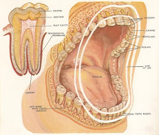
Human Teeth Anatomy Pics

Human Teeth Anatomy Pics:


Human Back Bone Anatomy Pics

Human Back Bone Anatomy Pics:


Human Heart Anatomy Pics

Human Heart Anatomy Pics:


Human Abdomen Anatomy Pics

Human Abdomen Anatomy Pics:


Human Head Anatomy Pics

Human Head Anatomy Pics


Tuesday, April 12, 2011

Causes, Symptoms Of Jaundice and Its Treatment

What is Jaundice?
Jaundice is not a disease but rather a sign that can occur in many different diseases.Jaundice, (also known as icterus) is a yellowish pigmentation of the skin, the conjunctival membranes over the sclerae (whites of the eyes), and other mucous membranes caused by hyperbilirubinemia (increased levels of bilirubin in the blood). Jaundice comes from the French word jaune, meaning yellow.The color of the skin and sclerae vary depending on the level of bilirubin. When the bilirubin level is mildly elevated, they are yellowish. When the bilirubin level is high, they tend to be brown.

Causes Of Jaundice:
Jaundice may be caused by several different disease processes. It is helpful to understand the different causes of jaundice by identifying the problems that disrupt the normal bilirubin metabolism and/or excretion.Hemoglobin present in the red blood cells is an important carrier of oxygen throughout the body. Our body replaces dead red blood cells with the new ones and these dead RBCs are processed by liver. This is a normal process, but when the RBCs die rapidly amount of bilirubin in blood increases very fast. Hence, increased amount of bilirubin in blood is one of the causes for jaundice. Other causes for jaundice are diseases like Malaria, Typhoid and Tuberculosis. Jaundice in infants is a serious condition where infection or abnormality in the functioning of liver may be probable causes for jaundice in new born.

Types Of Jaundice:
Jaundice types are categorized in accordance to their effects on liver:
a.    Pre-Hepatic
b.    Hepatocellular
c.    Post Hepatic
Pre-Hepatic:
               The pathology is occurring prior to the liver.In tropical countries, malaria can cause jaundice in this manner. Certain genetic diseases, such as sickle cell anemia, spherocytosis, thalassemia and glucose 6-phosphate dehydrogenase deficiency can lead to increased red cell lysis and therefore hemolytic jaundice. Commonly, diseases of the kidney, such as hemolytic uremic syndrome, can also lead to coloration.
Hepatocellular:
              The pathology is located within the liver.Hepaticellular (Hepatic) jaundice (in hepatocellular jaundice there is invariably cholestasis) can be caused by acute hepatitis, hepatotoxicity, and alcoholic liver disease, whereby cell necrosis reduces the liver's ability to metabolize and excrete bilirubin leading to a buildup of unconjugated bilirubin in the blood.Rat fever (leptospirosis) can also cause hepatic jaundice.
Post Hepatic:
             The pathology is located after the conjugation of bilirubin in the liver. Post-hepatic jaundice, also called obstructive jaundice, is caused by an interruption to the drainage of bile in the biliary system. The most common causes are gallstones in the common bile duct, and pancreatic cancer in the head of the pancreas. Also, a group of parasites known as "liver flukes" can live in the common bile duct,causing obstructive jaundice.

Symptoms Of Jaundice:
The first and foremost jaundice symptom is the skin color turning yellow. Tongue along with skin turns yellowish or pale. Patients feel extreme fatigue and weakness along with some common jaundice symptom like fever, headache and nausea.Jaundice symptom triggers pregnant women making them more prone to fever, vaginal bleeding, dizziness, frequent fainting episodes and abnormal thirst.

Treatment Of Jaundice:
Treatment depends on the underlying cause, and may involve removal of the offending agent; administration of antibiotic, antiviral, antifungal, or antiparasitic drugs; surgery to correct blockage; the use of chemotherapy, antiflammatory, or steroid medications, dietary changes; measures to minimize symptoms; or any combination of the above. The need to limit activity depends entirely on the underlying cause of the jaundice, but in general, only serious problems require severe limitations.


What causes cardiac arrest? and Treatment for cardiac arrest


Introduction to sudden cardiac arrest:
A natural disaster hits, the power goes off and the lights go out. It's a common scene that plays out during hurricane and tornado seasons, and it's very similar in trying to explain sudden cardiac arrest. The heart sustains an insult, the electricity is short circuited, the heart can't pump, and the body dies.

              The heart is an electrical pump, where the electricity is generated in special pacemaker cells in the upper chamber, or atrium, of the heart. This electrical spark is carried through pathways in the heart so that all the muscle cells contract at once and produce a heart beat. This pumps blood through the heart valves and into all the organs of the body so that they can do their work.

            This mechanism can break down in a variety of ways, but the final pathway in sudden death is the same: the electrical system is irritated and fails to produce electrical activity that causes the heart to beat. The heart muscle can't supply blood to the body, particularly the brain, and the body dies. Ventricular fibrillation (V Fib) is the most common reason for sudden death in patients. Without a coordinated electrical signal, the bottom chambers of the heart (ventricles) stop beating and instead, jiggle like Jello. Ventricular Fibrillation is treated with electrical shock, but for it to be effective, the shock usually needs to happen within less than four to six minutes, not only for it to be effective, but also to minimize brain damage from lack of blood and oxygen supply. Automatic external defibrillators (AEDs) are commonly available in public places to allow almost anybody to treat sudden death.

What is cardiac arrest?
Cardiac arrest is the sudden, abrupt loss of heart function. The victim may or may not have diagnosed heart disease. It's also called sudden cardiac arrest or unexpected cardiac arrest. Sudden death (also called sudden cardiac death) occurs within minutes after symptoms appear.A cardiac arrest is different from (but may be caused by) a heart attack, where blood flow to the muscle of the heart is impaired.

                Arrested blood circulation prevents delivery of oxygen to the body. Lack of oxygen to the brain causes loss of consciousness, which then results in abnormal or absent breathing. Brain injury is likely if cardiac arrest goes untreated for more than five minutes.For the best chance of survival and neurological recovery, immediate and decisive treatment is imperative.

What causes cardiac arrest?
The most common underlying reason for patients to die suddenly from cardiac arrest is coronary heart disease. Most cardiac arrests that lead to sudden death occur when the electrical impulses in the diseased heart become rapid (ventricular tachycardia) or chaotic (ventricular fibrillation) or both. This irregular heart rhythm (arrhythmia) causes the heart to suddenly stop beating. Some cardiac arrests are due to extreme slowing of the heart. This is called bradycardia.

Other factors besides heart disease and heart attack can cause cardiac arrest. They include respiratory arrest, electrocution, drowning, choking and trauma. Cardiac arrest can also occur without any known cause.

What Are the Symptoms of Sudden Cardiac Arrest?
Some people may experience symptoms of sudden cardiac arrest, such as a racing heartbeat or they may feel dizzy, alerting them that a potentially dangerous heart rhythm problem has started. In over half of the cases, however, sudden cardiac arrest occurs without prior symptoms.
However, due to inadequate cerebral perfusion, the patient will be unconscious and will have stopped breathing. The main diagnostic criterion to diagnose a cardiac arrest, (as opposed to respiratory arrest which shares many of the same features), is lack of circulation, however there are a number of ways of determining this.

Diagnosis:
Cardiac arrest is synonymous with clinical death.A cardiac arrest is usually diagnosed clinically by the absence of a pulse. In many cases lack of carotid pulse is the gold standard for diagnosing cardiac arrest, but lack of a pulse (particularly in the peripheral pulses) may be a result of other conditions (e.g. shock), or simply an error on the part of the rescuer. Studies have shown that rescuers often make a mistake when checking the carotid pulse in an emergency, whether they are healthcare professionals or lay persons.
                 Owing to the inaccuracy in this method of diagnosis, some bodies such as the European Resuscitation Council (ERC) have de-emphasised its importance. The Resuscitation Council (UK), in line with the ERC's recommendations and those of the American Heart Association,have suggested that the technique should be used only by healthcare professionals with specific training and expertise, and even then that it should be viewed in conjunction with other indicators such as agonal respiration.

Prevention:
 
With positive outcomes following cardiac arrest unlikely, an effort has been spent in finding effective strategies to prevent cardiac arrest. With the prime causes of cardiac arrest being ischemic heart disease, efforts to promote a healthy diet, exercise, and smoking cessation are important. For people at risk of heart disease, measures such as blood pressure control, cholesterol lowering, and other medico-therapeutic interventions are used.

What is the treatment for sudden cardiac arrest?
The vast majority of people whose heart stops beating unexpectedly have ventricular fibrillation. The definitive treatment for this is defibrillation using electricity to shock the heart back into a regular rhythm. With technological advances, AEDs are now a routine sight wherever people congregate.

              Communities which institute public CPR education, use of AEDs, and rapid activation of 911 emergency medical services have dramatically increased survival rates from sudden cardiac arrest. Unfortunately, because the brain is so sensitive to the lack of oxygen and blood flow, unless treatment occurs within four to six minutes, there is a high risk of some permanent brain damage.

           Should the patient survive to be transported to the hospital, the reason for collapse and sudden death will need to be diagnosed. Regardless, the ABCs of resuscitation will be re-evaluated. Airway, Breathing, and Circulation (heart beat and blood pressure) will be supported, and admission to an intensive care unit is most likely.

Unwanted Hair Removal Tips


The desire to remove excess or dark hair usually begins in adolescence and seems to continue until the day we die. Whether it is hair on the face, armpits, legs, bikini line, or other body parts, many women AND men are intent upon having the hair on their scalp be the only visible hair on their bodies.As the people get older or even at some point in hormonal changes in men and women, sometimes hair do appear in the unusual areas of our body. When you want a quick removal of unwanted hair, here are pointers that can help make it as trouble-free as it can be.
Bikini line:
Use hair removal cream, razors or tweezers, these can all work efficiently in this areas. Just be sure not to cut-off hair from delicate areas. Another option is laser treatment.These laser treatments are always performed by a licensed clinician or M.D. Electrolysis removes unwanted hairs with an electric needle that probes into single hairs (one at a time), conducting an electrical current that kills the root and prevents regrowth. Laser hair removal uses the pulsation of a laser light to kill hair growth when applied to small areas (about the size of a quarter) around unwanted hairs.
Lip area :
For lip hair removal there are a number of options that are available.Due to the small area that is generally covered by the lip, the area to be treated is obviously not as big as other common areas with unwanted hair like the arms, legs or back.Shaving is generally not recommended for lip hair removal (unless you're a guy who has to shave daily anyways!) since it is a very temporary method and you'll just have to regularly shave to keep the hair-free look.Popular options available for lip hair removal include laser hair removal,pulsed lLight hair removal,waxing hair removal and tweezing hair removal etc.
Nose hair:
Because of the location of the hairs which is inside the nose, some of the conventional methods of hair removal are not applicable to it. Waxing, shaving and the use of depilatories can prove to be very difficult when it comes to removing these very tiny hairs. These methods which are capable of removing much hair all at once are not that popular among people who want to have their nose hair removed.When tips for unwanted nasal hair removal are desired, it is important to exercise caution. Hair in the nasal passage is there for a reason. It helps filter the air people breathe. With this in mind, it's best to not tweeze or otherwise pull out nasal hair. It can, however, be trimmed. The best method for this is to use specialized nose hair clippers. These are designed to make access into the nasal cavity easier.
Ear hair:
Men in particular can find some rather odd hairs growing out of their ears as they age. While this hair is not quite as important as nasal hair, it should not be completely removed when in the ear canal. Trim instead. For the outer ear, use tweezers to pull hair out from the root if at all possible. Do be careful with hair removal even on the outside, as it can be a bit more painful than many realize.


Monday, April 11, 2011

Natural Remedies For Hair Growth

Hair loss is a metabolic disorder where hair starts thinning, falling, or disappearing. The papilla which is an upgrowth at the base of the follicle, actually produces hair when a special group of cells turn amino acids into keratin, a type of protein of which hair is made.Although many experts say there is no way to accelerate the growth of hair, there are several home remedies that people have used and passed down from generation to generation asserting that they do make hair grow faster.
Onion:
Take a fresh onion. Get the juice of the onion by grinding it in  mixer.Apply the juice directly on the scalp.Leave it for 1/2 and hour.Wash your hair with shampoo.This will help in restoring the hair follicles and helps in hair growth.
Grapeseed oil:
Grape seed Oil promotes hair growth.Test found that antioxandants present in grape seeds promote proliferation of hair follicles cells and that it may be used to induce hair growth. New Studys show that grape seed oil is excellent to use in scalp oil to prevent hair loss.
Aloe Vera Shampoo:
Aloe vera shampoo has been around for a while, there are aloe vera conditioners too. It is very good for your hair, and you will get a nice shine and improved hair health with daily use. Because aloe vera is so beneficial, with vitamins, amino acids and other nutrients, it helps to increase the vitality of our hair. Hair strands are porous and the aloe vera is absorbed into the hair shaft and increases the vitality of your hair. 
Energetic breakfast:
1 tablespoon of wheat germ, 1 tablespoon of brewer’s yeast, 1 tablespoon soy lecithin, 1 tablespoon honey, and one cup of yogurt.Home-remedies-site.com Combine all ingredients in the yogurt and eat it every day before breakfast. In one week you will see results; a hair full of strength and growing faster.
Nutrition for your hair:
The effects of B-Vitamins like Biotin (B7) and Niacin (B3) on hair growth are well documented. Consuming foods with healthy concentrations of B-Vitamins (such as lean meats, green vegetables and whole grains) improves metabolism, digestion and blood circulation – all of which have noticeable impacts on hair growth. Also, foods that are rich in Vitamins C and E can increase energy levels and drastically improve the appearance of hair and skin.

Thursday, April 7, 2011

Causes of Beriberi,Symptoms and Treatment


What Is Beriberi:
Beriberi is a disease in which the body does not have enough thiamine.beriberi, nutritional disorder caused by a deficiency of thiamin (vitamin B1) and characterized by impairment of the nerves and heart. General symptoms include loss of appetite and overall lassitude, digestive irregularities, and a feeling of numbness and weakness in the limbs and extremities.(The term beriberi is derived from the Sinhalese word meaning “extreme weakness.”) In the form known as dry beriberi, there is a gradual degeneration of the long nerves, first of the legs and then of the arms, with associated atrophy of muscle and loss of reflexes.
                    In wet beriberi, a more acute form, there is edema (overabundance of fluid in the tissues) resulting largely from cardiac failure and poor circulation.In infants breast-fed by mothers who are deficient in thiamin, beriberi may lead to rapidly progressive heart failure.
                  Probably the most important factor in the reduction of beriberi is the general increase in overall food consumption so that the staple diet is varied and contains legumes and pulses, which contain a large amount of thiamin. There are two main clinical types of beriberi, which, surprisingly, only rarely occur together.

History:
 
In Asia, where polished white rice was the common staple food of the middle class, beriberi resulting from lack of vitamin B1 was endemic. In 1884, Takaki Kanehiro, a British-trained Japanese medical doctor of the Japanese Navy, observed that beriberi was endemic among low-ranking crew who often ate nothing but rice, but not among crews of Western navies and officers who consumed a Western-style diet.In 1883, Kanehiro learned of a very high incidence of beriberi among cadets on a training mission from Japan to Hawaii, via New Zealand and South America that lasted for 9 months. On board, 169 men out of 376 developed the disease and 25 died. With the support of the Japanese Navy, he conducted an experiment in which another ship was deployed on the same route and under identical conditions, except that its crew was fed a diet of meat, fish, barley, rice, and beans.At the end of the voyage, this crew had suffered only 14 cases of beriberi and no deaths.
                      This convinced Kanehiro and the Japanese Navy that diet was the cause of beriberi.This was confirmed in 1897, when Dr. Christiaan Eijkman, a Dutch physician and pathologist, demonstrated that beriberi is caused by poor diet. He discovered that feeding unpolished rice instead of the polished variety to chickens helped to prevent beriberi in the chickens.The following year, Sir Frederick Hopkins postulated that some foods contained "accessory factors"—in addition to proteins, carbohydrates, fats, and salt—that were necessary for the functions of the human body.
                        In 1901, Gerrit Grijns (May 28, 1865 – November 11, 1944), Dutch physician and assistant to Christiaan Eijkman in Netherlands Indies, correctly interpreted the disease as a deficiency syndrome.Indeed, it was later shown that beriberi results from the deficiency of thiamine (vitamin B1).

symptoms: 
1)Appetite loss
2)Tiredness
3)Irritability
4)Muscle aches
5)Limb pains
6)Swollen joints
7)Hand paralysis
8)Foot paralysis
9)Heart problems

Causes:
Beriberi is caused by a lack of thiamine (vitamin B1). Thiamine occurs naturally in unrefined cereals and fresh foods, particularly whole grain bread, fresh meat, legumes, green vegetables, fruit, milk, etc. Beriberi is therefore common in people whose diet excludes these particular types of nutrition e.g. as a result of famine.
                        Beriberi may be found in people whose diet consists mainly of polished white rice, which is very low in thiamine because the thiamin-bearing husk has been removed. It can also be seen in chronic alcoholics (Wernicke-Korsakoff syndrome), Arsenic poisoning causes alterations in cellular metabolism resulting in blockage of thiamine use which results in thiamine deficiency without any dietary shortfall.The mechanism of arsenic neuropathy may be similar to the neuropathy of thiamine deficiency [Sexton and Gowdy 1963], whereby arsenic inhibits the conversion of pyruvate to acetyl coenzyme A and thus blocks the Krebs cycle.The disease was often found in Asian countries (especially in the 19th century and before), due to those countries' reliance on white rice as a staple food.


Treatment: 
The goal of treatment is to provide the thiamine the body is lacking. This is done with thiamine supplements which are given by injection or taken by mouth.Other vitamins may also be recommended.
                   Subsequent blood tests will determine if the thiamine supplements are being effective.Treatment for beriberi is with thiamine hydrochloride, either in tablet form or injection. A rapid and dramatic recovery within hours can be made when this is administered to patients, and their health can be improved within an hour of starting treatment.In emergency situations where concentrated thiamin supplements are unavailable, feeding the patient with a thiamin-rich diet (e.g. whole grain brown bread) will lead to recovery, though at a much slower rate.

Prevention:
Eating a proper diet that is rich in thiamine and other vitamins will prevent beriberi.Nursing mothers should make sure that their diet contains all vitamins and be sure that infant formulas contain thiamine.

People who drink heavily should try to cut down or quit, and take B vitamins to make sure their body is properly absorbing and storing thiamine.

Monday, April 4, 2011

causes of Appendicitis and Treatment

 What is appendicitis:
Appendicitis is a painful swelling and infection of the appendix.Appendicitis is the inflammation of the appendix.The symptoms are pain in the belly button area which in the end will move in the right side of the lower abdomen and will be much worse, low graded fever, nauseas and sometimes even vomiting, loss of appetite and constipation or diarrhea.
Some people might not have all these symptoms and some might not have any of them, but still have appendicitis.The appendix is a closed, narrow tube up to several centimeters in length that attaches to the cecum (the first part of the colon) like a worm.
(The anatomical name for the plant, ie vermiform, worm-like appendage.) The inner lining of the plant produces a small amount of mucus is flowing through the open center of the plant and in the appendix.The wall of the appendix contains lymphatic tissue that is part of the immune system to produce antibodies. As theRest of the colon, the wall of the annex includes a layer of muscles, but the muscle is poorly developed.

What is the appendix:
The appendix is a fingerlike pouch attached to the large intestine and located in the lower right area of the abdomen. Scientists are not sure what the appendix does, if anything, but

removing it does not appear to affect a person’s health.The inside of the appendix is called the appendiceal lumen. Mucus created by the appendix travels through the appendiceal lumen and empties into the large intestine.
  The inner lining of the appendix produces a small amount of mucus that flows through the open center of the appendix and into the cecum. The wall of the appendix contains lymphatic tissue that is part of the immune system for making antibodies.Like the rest of the colon, the wall of the appendix also contains a layer of muscle, but the layer of muscle is poorly developed.

What causes appendicitis:
Obstruction of the appendiceal lumen causes appendicitis. Mucus backs up in the appendiceal lumen, causing bacteria that normally live inside the appendix to multiply. As a result, the appendix swells and becomes infected. Sources of obstruction include
1)feces, parasites, or growths that clog the appendiceal lumen
2)enlarged lymph tissue in the wall of the appendix, caused by infection in the gastrointestinal tract or elsewhere in the body
3)inflammatory bowel disease, including Crohn’s disease and ulcerative colitis
4)trauma to the abdomen
An inflamed appendix will likely burst if not removed. Bursting spreads infection throughout the abdomen—a potentially dangerous condition called peritonitis.

What are the symptoms of appendicitis:
Most people with appendicitis have classic symptoms that a doctor can easily identify. The main symptom of appendicitis is abdominal pain.
The abdominal pain usually
1)occurs suddenly, often causing a person to wake up at night
2)occurs before other symptoms
3)begins near the belly button and then moves lower and to the right
4)is new and unlike any pain felt before
5)gets worse in a matter of hours
6)gets worse when moving around, taking deep breaths, coughing, or sneezing
Other symptoms of appendicitis may include:
1)loss of appetite
2)nausea
3)vomiting
4)constipation or diarrhea
5)inability to pass gas
6)a low-grade fever that follows other symptoms
7)abdominal swelling
8)the feeling that passing stool will relieve discomfort
Symptoms vary and can mimic other sources of abdominal pain, including
1)intestinal obstruction
2)inflammatory bowel disease
3)pelvic inflammatory disease and other gynecological disorders
4)intestinal adhesions
5)constipation

Diagnosis:
Diagnosis is based on patient history (symptoms) and physical examination backed by an elevation of neutrophilic white blood cells. Histories fall into two categories, typical and atypical. Typical appendicitis usually includes abdominal pain beginning in the region of the umbilicus for several hours, associated with anorexia, nausea or vomiting. The pain then "settles" into the right lower quadrant, where tenderness develops.
Atypical histories lack this typical progression and may include pain in the right lower quadrant as an initial symptom. Atypical histories often require imaging with ultrasound and/or CT scanning.
A pregnancy test is vital in all women of child bearing age, as ectopic pregnancies and appendicitis present with similar symptoms. The consequences of missing an ectopic pregnancy are serious, and potentially life threatening. Furthermore the general principles of approaching abdominal pain in women (in so much that it is different from the approach in men) should be appreciated.Blood Test Most patients suspected of having appendicitis would be asked to do a blood test. 50% of the time, the blood test may be normal, so it is not fool proof in diagnosing appendicitis.

How is appendicitis treated:
Surgery:
Typically, appendicitis is treated by removing the appendix. If appendicitis is suspected, a doctor will often suggest surgery without conducting extensive diagnostic testing. Prompt surgery decreases the likelihood the appendix will burst.

Surgery to remove the appendix is called appendectomy and can be done two ways. The older method, called laparotomy, removes the appendix through a single incision in the lower right area of the abdomen.
The newer method, called laparoscopic surgery, uses several smaller incisions and special surgical tools fed through the incisions to remove the appendix. Laparoscopic surgery leads to fewer complications, such as hospital-related infections, and has a shorter recovery time.Surgery occasionally reveals a normal appendix. In such cases, many surgeons will remove the healthy appendix to eliminate the future possibility of appendicitis. Occasionally, surgery reveals a different problem, which may also be corrected during surgery.

Sometimes an abscess forms around a burst appendix—called an appendiceal abscess. An abscess is a pus-filled mass that results from the body’s attempt to keep an infection from spreading. An abscess may be addressed during surgery or, more commonly, drained before surgery.
To drain an abscess, a tube is placed in the abscess through the abdominal wall. CT is used to help find the abscess. The drainage tube is left in place for about 2 weeks while antibiotics are given to treat infection.
Six to 8 weeks later, when infection and inflammation are under control, surgery is performed to remove what remains of the burst appendix.

Nonsurgical Treatment:
Nonsurgical treatment may be used if surgery is not available, if a person is not well enough to undergo surgery, or if the diagnosis is unclear. Some research suggests that appendicitis can get better without surgery.
Nonsurgical treatment includes antibiotics to treat infection and a liquid or soft diet until the infection subsides. A soft diet is low in fiber and easily breaks down in the gastrointestinal tract.

Recovery:
With adequate care, most people recover from appendicitis and do not need to make changes to diet, exercise, or lifestyle. Full recovery from surgery takes about 4 to 6 weeks. Limiting physical activity during this time allows tissues to heal.

what is Alcoholic Hepatitis and Treatment?


what is alcoholic hepatitis:

Alcoholic hepatitis is hepatitis (inflammation of the liver) due to excessive intake of alcohol. While distinct from cirrhosis, it is regarded as the earliest stage of alcoholic liver disease.
Symptoms are jaundice, ascites (fluid accumulation in the abdominal cavity), fatigue and hepatic encephalopathy (brain dysfunction due to liver failure).Mild cases are self-limiting, but severe cases have a high risk of death. Severe cases may be treated with glucocorticoids.
This will depend on the severity of the alcoholic hepatitis.In mild cases only abstinence from alcohol and nutritional support are required.But in acute severe alcoholic hepatitis – characterised by jaundice, easy bruising, abnormal blood tests and sometimes the presence of extra fluid within the abdomen (ascites) – hospital admission is necessary.Steroids can also help some of these patients, but even with steroids 10 to 15 per cent die within three months of developing the disease.Kidney failure, if it occurs, leads to an almost 100 per cent mortality rate.
Symptoms:
1)Anorexia
2)Nausea and Vomiting
3)Weight loss
4)Fever
5)Generalized Abdominal Pain
6) Jaundice is usually one of the most common symptom. Some common mode of presentations are: jaundice - 50% of the patients, ascites in 30 - 60% and splenomegaly - 15% of the patients.
Alcoholic hepatitis is distinct from cirrhosis caused by long-term alcohol consumption. Alcoholic hepatitis can occur in patients with chronic alcoholic liver disease and alcoholic cirrhosis.
Alcoholic hepatitis by itself does not lead to cirrhosis, but cirrhosis is more common in patients with long term alcohol consumption.
Signs:
1)Tender Hepatomegaly (80-90% of cases)
2)aundice
3)Ascites
4)Splenomegaly
5)Spider angioma

  Some alcoholics develop acute hepatitis as an inflammatory reaction to the cells affected by fatty change. This is not directly related to the dose of alcohol. Some people seem more prone to this reaction than others. This is called alcoholic steatonecrosis and the inflammation probably predisposes to liver fibrosis.Features of portal hypertension - Ascites, dilated abdominal wall veins, splenomegaly and oesophageal varices are not a prominent features of pure alcoholic hepatitis.Liver is very large and tender on palpation its surface is smooth and consistency is soft to firm.
Diagnosis:
The ratio of aspartate aminotransferase to alanine aminotransferase is usually 2 or more.In most cases, the liver enzymes do not exceed 500.
Fatty liver is usually diagnosed in the asymptomatic patient who is undergoing evaluation for abnormal liver function tests; typically, aminotransferase levels are less than twice the upper limit of normal. No laboratory test is diagnostic of fatty liver.
Characteristic ultrasonographic findings include a hyperechoic liver with or without hepatomegaly. Liver biopsy is rarely needed to diagnose fatty liver in the appropriate clinical setting, but it may be useful in excluding steatohepatitis or fibrosis.
Treatment:
Clinical practice guidelines by the American College of Gastroenterology recommend corticosteroids.Patients should be risk stratified using a MELD Score or Child-Pugh score.
Hospitalization is indicated to expedite a diagnostic evaluation of patients with jaundice, encephalopathy, or ascites of unknown cause. In addition, patients with known alcoholic liver disease who present with renal failure, fever, inadequate oral intake to maintain hydration, or rapidly deteriorating liver function, as demonstrated by progressive encephalopathy or coagulopathy, should be hospitalized.

Causes of Tuberculosis and Treatment

Introduction about tuberculosis: 
In an age when  we believe that we have the tools to conquer most diseases,the ancient scourge of tuberculosis(tb) still causes 2 million deaths a year worldwide-more than any other single infections organism-remainding us that we still have a long way to go.Even equipped with drugs to treat TB effectively,we haven't managed to eradicate this deadly infection.
From where this disease is effected:
         Mycobacterium tuberculosis, the bacteria that causes tuberculosis, has been around for centuries. Recently, fragments of the spinal columns from Egyptian mummies from 2400 B.C.E. were found to have definite signs of the ravages of this terrible disease. Also called consumption, TB was identified as the most widespread disease in ancient Greece, where it was almost always fatal.
        But it wasn't until centuries later that the first descriptions of the disease began to appear. Starting in the late seventeenth century, physicians began to identify changes in the lungs common in all consumptive, or TB, patients. At the same time, the earliest references to the fact that the disease was infectious began to appear.
        In 1720, the English doctor Benjamin Marten was the first to state that TB could be caused by “wonderfully minute living creatures.” He went further to say that it was likely that ongoing contact with a consumptive patient could cause a healthy person to get sick. Although Marten's findings didn't help to cure TB, they did help people to better understand the disease. The sanitorium, which was introduced in the mid-nineteenth century, was the first positive step to contain TB. Hermann Brehmer, a Silesian botany student who had TB, was told by his doctor to find a healthy climate. He moved to the Himalayas and continued his studies. He survived his bout with the illness, and after he received his doctorate, built an institution in Gorbersdorf, where TB patients could come to recuperate.
        They received good nutrition and were outside in fresh air most of the day. This became the model for the development of sanitoria around the world.In 1865, French military doctor Jean-Antoine Villemin demonstrated that TB could be passed from people to cattle and from cattle to rabbits. In 1882, Robert Koch discovered a staining technique that allowed him to see the bacteria that cause TB under a microscope.

        Until the introduction of surgical techniques to remove infected tissue and the development of x-rays to monitor the disease, doctors didn't have great tools to treat TB. TB patients could be isolated, which helped reduce the spread of the disease, but treating it remained a challenge.
Transmission of disease:
       Some diseases, such as influenza, are contagious or infectious, and can be transmitted by any of a variety of mechanisms, including coughs and sneezes, sexual transmission, by bites of insects or other carriers of the disease, from contaminated water or food, and so forth.Other diseases, such as cancer and heart disease, are not considered to be due to infection, although microorganisms may play a role.
Latent TB Infection:
Pulmonary tuberculosis, or TB of the lungs, is the most common form of TB. TB can also attack the spine, bones and joints, the central nervous system, the gastrointestinal tract, the lymph system, and the heart.

            Only 5 to 10 percent of healthy people who come in contact with TB bacteria will ever get sick. The vast majority of them will live with dormant TB bacteria in their bodies throughout their lives, because their immune systems are able to fight the bacteria and stop them from growing. People with latent TB don't feel sick, don't have symptoms, and can't spread TB. However, the bacteria remain alive in the body and can become active later. They have what are called latent infections.

          TB is spread through the air, not through handshakes, sitting on toilet seats, or sharing dishes and utensils with someone who has TB. However, casual exposure is not sufficient for someone to get TB.

          If at some point in their lives their immune system is weakened, the once-dormant bacteria may begin to grow again and cause active tuberculosis. Sometimes, doctors will recommend that people with latent TB infections take medicine to prevent development of active disease. The medicine is usually a drug called isoniazid (INH), which kills the TB bacteria that are in the body. Usually the course of treatment is six to nine months. Children and people with HIV infection, however, sometimes have to take INH for a longer period of time.
symptoms:
1)A bad cough that lasts longer than two weeks.
2)Pain in the chest.
3)Coughing up blood or sputum.
4)Weakness or fatigue.
5)Weight loss.
6)Loss of appetite.
7)Chills.
8)Fever.
9)Sweating at night.
Treating TB:
        Most of the time TB can be cured with antibiotics. If you have TB, you will need to take several drugs. This is because there are many bacteria to be killed. Taking multiple drugs also helps to prevent the bacteria from becoming drug resistant and, thus, much more difficult to cure.

        If you have TB of the lungs, or pulmonary TB, you are probably infectious. This means that you can spread the disease by coughing or sneezing. Fortunately, after a couple of weeks of taking medicine, most people are no longer infectious and they begin to feel better. Usually they can return to life as usual. But that doesn't mean all the bacteria are killed. People often have to take TB medicine for six to nine months before all the bacteria are killed.
Why Is It Important to Take TB Medicine for So Long?
        TB bacteria die very slowly. Even when patients start to feel better, the bacteria are alive in their bodies. They have to keep taking medicine until all the bacteria are dead, otherwise they can get sick again and infect others.

       Another danger of not completing the whole course of therapy is the rise of drug-resistant TB. If you stop taking your medicine and some of the bugs are still alive, they may become resistant to the drugs you were taking, so that if you get sick again, you will need different drugs to kill the bacteria because the old ones won't work. These additional drugs, called second-line drugs, must be taken for a very long time, sometimes up to two years, and their side effects can be quite serious.

       The only way to get better is to take your medicine as prescribed by the doctor. Most public health officials advocate Directly Observed Therapy (DOTS), which is when a health-care worker meets with the patient every day, or several times a week, to be sure they take their medicine. Sometimes they meet at the patient's home or at a hospital or TB clinic. Some DOTS programs provide medicine that can be taken only two or three times a week instead of every day. In addition to ensuring that the patient takes their medication as prescribed, the health care worker also monitors side effects.

       DOTS works and it is used in many countries. It is the World Health Organization's recommended method for successfully treating TB.Patients with active TB who have to go to the hospital may be put in special rooms with negative air pressure. This keeps TB from spreading from room to room, or out into hospital hallways. People who enter the rooms will wear special facemasks to protect themselves.

Social significance of disease:
      The identification of a condition as a disease, rather than as simply a variation of human structure or function, can have significant social or economic implications. The controversial recognitions as diseases of post-traumatic stress disorder, also known as "shell shock"; repetitive motion injury or repetitive stress injury (RSI); and Gulf War syndrome have had a number of positive and negative effects on the financial and other responsibilities of governments, corporations, and institutions towards individuals, as well as on the individuals themselves.
Multi-Drug-Resistant TB (MDR-TB):
    When TB patients don't take their medicine properly, the TB bacteria may become resistant to certain drugs. This means the drugs can no longer kill the bacteria. Drug resistance is most likely to occur when people …
Have spent time with someone with drug-resistant TB.
Don't take their medicine regularly.
Don't take all their medicine.
Develop TB after they've taken TB drugs before.
Come from areas where drug-resistant TB is common.
      Multi-drug-resistant TB (MDR-TB) is a form of tuberculosis that is resistant to two or more of the first-line drugs used to treat the disease. When the bacteria resist the antibiotics used to attack them, they relay that ability to new bacteria that is produced. People with multi-drug-resistant TB must be treated with special second-line drugs. These drugs don't kill the bacteria as well as the first-line drugs, and they often cause more severe side effects.

         If a person with MDR-TB spreads the disease to someone else and that person comes down with active disease, it will be multi-drug-resistant from the beginning. In the early 1990s, there were several outbreaks of multi-drug-resistant TB in New York City hospitals that were caused primarily by the spread of one strain, strain W, that went from patient to patient to patient. This strain was resistant to between seven and nine drugs. A large number of these patients died, and many health-care workers now have latent infections with this highly resistant strain.